
Consolidating Multi-Site Imaging Data: How Methods Compare
No single preprocessing step can erase scanner differences. Consolidating multi-site imaging data calls for a combination of consistent acquisition protocols, well-organized metadata, and a harmonization method suited to the images and analysis. Standardizing a DTI protocol is a useful first step, but it doesn’t eliminate systematic differences between scanners.
It’s easy to assume that matching file formats or using the same correction method will make scans directly comparable. In practice, differences between sites and scanners can remain, and harmonization can affect how well findings generalize to new data. This article explains where inter-scanner variability comes from and compares protocol standardization, statistical harmonization, and image-level approaches. You’ll also learn how to weigh compatibility, preservation of meaningful biological differences, and performance across sites when planning a multi-site imaging workflow.
Key Takeaways
- Use DICOM and FHIR for complementary parts of interoperability: imaging data exchange and clinical information sharing.
- Align acquisition protocols and metadata across sites, then choose harmonization methods that fit the data and analysis goals.
- Compare centralized, federated, and hybrid architectures by weighing data access, governance, and operational needs.
- Match records with privacy safeguards and clear identity-management processes to reduce the risk of misidentification.
- Build privacy, consent, and data-sharing requirements into the workflow from the start, not as a final data-transfer step.
What data standards support interoperability across imaging sites, such as DICOM and FHIR?
DICOM carries medical images and the metadata needed to interpret them. A CT or MRI study includes image instances, series details, and identifiers that help systems organize related files. DICOM also defines how imaging systems exchange, store, and retrieve these objects. This common structure lets a receiving archive process images from different scanner vendors without requiring identical equipment. It does not, by itself, make acquisition protocols or image measurements equivalent.
DICOMweb provides familiar web-based services for working with DICOM objects. QIDO-RS searches for studies, series, or instances; WADO-RS retrieves them; and STOW-RS stores them. These functions support workflows where a site needs to find and transfer imaging data without moving an entire archive. DICOM identifiers, including study and series instance UIDs, help preserve relationships among files. Keep those identifiers intact during export and import, and verify that each image remains linked to the correct study.
FHIR serves a different role. Its structured resources represent clinical information exchanged between health systems. In imaging workflows, the ImagingStudy resource can describe a study and reference its DICOM series or instances. A ServiceRequest can represent an imaging order, while a DiagnosticReport can carry the resulting interpretation. FHIR helps connect images to their clinical context, but it does not replace DICOM as the format for storing diagnostic image pixels.
Use these standards as complementary layers. For example, a FHIR ImagingStudy record can point to a DICOM MRI study, while a DiagnosticReport carries the radiologist’s findings. An integration can then map patient, order, accession, and study identifiers between systems. Preserve original identifiers alongside any local values. This mapping helps prevent an order or report from becoming detached from its images during exchange.
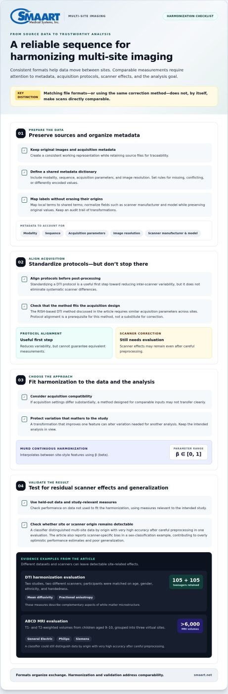
Standard formats still leave scanner effects unresolved. In one DTI harmonization evaluation, researchers compared two studies acquired using two different scanners. They retained 105 teenagers from each study after matching age, gender, ethnicity, and handedness. Mean diffusivity and fractional anisotropy described complementary aspects of white matter microstructure. DICOM can carry these images and metadata, but file compatibility alone cannot ensure those measurements are comparable across sites.
A separate evaluation used more than 6,000 T1- and T2-weighted MRI volumes from children aged 9-10 in the ABCD study. The scans came from General Electric, Philips, and Siemens scanners, grouped into three virtual sites. A classifier could still distinguish multi-site brain imaging data by origin with very high accuracy after careful preprocessing. For consolidating multi-site imaging data, use DICOM and FHIR to structure exchange, then assess scanner and protocol effects separately.
MURD’s continuous harmonization interpolates between site-style features using a parameter β ranging from [0, 1]. The parameter represents a position along that interpolation between site-style features.
How should imaging data from different sites be harmonized for formats, metadata, and protocols?
Start by preserving each site’s original images and acquisition metadata. Create a consistent working representation, but retain the source files for traceability. DICOM studies can differ in how fields are populated, even when the images use the same format. Map local labels to shared terms and record the mapping rules. For example, normalize scanner manufacturer and model fields without overwriting their original values. This lets analysts compare records and trace unexpected results back to their source.
Next, define a metadata dictionary before combining datasets. Include fields that affect interpretation, such as modality, sequence, acquisition parameters, and image resolution. Set rules for missing, conflicting, or differently encoded values. A text label used at one site may need to be mapped to a consistent coded term elsewhere. Keep an audit trail of every transformation. This makes quality checks repeatable and helps distinguish biological variation from inconsistent documentation.
Align acquisition protocols before relying on post-processing. Standardizing a DTI protocol is a first step toward reducing inter-scanner variability in multi-site studies, but it does not eliminate systematic scanner differences. A DTI harmonization evaluation compared two studies acquired using two different scanners. Its results illustrate why matching protocol descriptions alone cannot guarantee equivalent measurements. Record acquisition details that shape the signal, then assess scanner effects in the resulting data.
Choose a harmonization method that fits the acquisition design. The RISH-based DTI method discussed in the study requires similar acquisition parameters across sites. Protocol alignment is therefore a prerequisite, not a substitute for correction. If acquisition settings differ substantially, a method designed for comparable inputs may not transfer cleanly. Keep the intended analysis in view, too: a transformation that improves one feature can alter variation needed for another analysis.
Validate harmonization using held-out data and measures relevant to the study. The research found that current approaches did not remove scanner-specific bias in a sex-classification example. That bias contributed to overly optimistic performance estimates and poor generalization. Test whether models can still identify the site or scanner from harmonized data, then assess whether meaningful clinical or research signals remain. A reduction in site effects alone does not prove success.
Use a documented workflow for each dataset:
- Inventory formats, metadata, and protocol differences before transformation.
- Map fields and units while preserving the original values.
- Apply harmonization only where its acquisition assumptions fit.
- Evaluate results on separate data and record limitations.
Multi-site harmonization remains an open challenge, particularly when advanced machine-learning methods are used for predictive modeling. A clear protocol, metadata map, and validation plan make the process easier to review and reproduce.
What are the best platforms and architectures for consolidating multi-site imaging data?
The best architecture depends on where images must reside, who needs access, and how sites govern data. A centralized model moves imaging from each location into a shared archive, such as a vendor-neutral archive (VNA) or picture archiving and communication system (PACS). It gives authorized teams one place to search studies and apply common workflows. It also concentrates storage, access controls, and system dependencies, making governance and continuity planning important.
A federated architecture keeps images at their originating sites. A shared index or broker routes queries to local PACS archives and returns results to authorized users. This can reduce unnecessary transfers and preserve local control. It also relies on dependable network connections, compatible query interfaces, and consistent identity mapping. For example, a radiologist searching across hospitals needs a common way to locate studies, even when each hospital maintains its own archive.
A hybrid model combines both approaches. Sites may retain operational copies locally while sending selected studies or metadata to a central repository. A research network, for instance, could centralize approved datasets while clinical teams continue using local PACS. This can support different access needs, but it adds synchronization tasks. Before deployment, define which system owns each record, how updates propagate, and how duplicate studies are handled.
Cloud hosting describes where services run, not the complete architecture. A cloud PACS or VNA can support centralized access, while cloud storage can also connect to federated site systems. Evaluate how the platform handles DICOM objects, DICOMweb interfaces, metadata search, and integration with electronic medical record systems. FHIR can link clinical resources to imaging records, but it does not store image pixels. Keep this distinction clear when matching platform capabilities to workflow requirements.
Compare architectures against practical operating needs, not feature lists alone. A site with limited network capacity may prioritize local access and selective transfer. A distributed research team may need a shared catalog, controlled access, and traceable dataset preparation. Map the workflow from image creation through retrieval, review, sharing, and retention. Then identify where each platform stores images, metadata, and related clinical context.
- Centralized: Choose a shared PACS or VNA when unified search and central administration are priorities.
- Federated: Keep site archives in place when local custody matters and cross-site discovery can work through shared services.
- Hybrid: Combine local operations with central access when clinical and research workflows have different needs.
- Cloud-hosted: Assess access, integration, storage governance, and continuity as part of the architecture, not as isolated features.
Before selecting a platform, document data ownership, access roles, identifier handling, and transfer rules. Test a representative workflow across sites, including image retrieval and metadata search. Technical compatibility alone is not enough: the platform also needs clear governance and reliable integrations. Those decisions determine whether a consolidated environment supports daily imaging operations as well as cross-site analysis.
How can patient identities and records be matched across imaging sites safely
Start with a defined identity-matching workflow, not a single field. A medical record number (MRN) may identify a patient within one hospital, while another site assigns a different MRN. DICOM fields such as PatientID, PatientName, and PatientBirthDate can help locate candidate records, but they are not proof of identity on their own. Document which identifiers each site supplies and how the matching process uses them. This groundwork is essential when consolidating multi-site imaging data because local identifiers rarely function as shared keys.
Use a trusted identity service, such as an enterprise master patient index, to connect records across organizations. The index can maintain cross-references between local identifiers and a shared enterprise identity. For example, Site A’s MRN and Site B’s MRN can point to one matched patient record without replacing either source value. Keep the relationship traceable so authorized staff can review how the records were linked.
Combine exact matching with exception handling. A deterministic rule may match records when agreed fields align, such as a verified enterprise identifier. A probabilistic process can compare several attributes when local records differ, including name spelling, date of birth, or address. Treat close matches as candidates for review rather than automatic merges. Otherwise, a mistyped birth date or name change could falsely link two people.
Apply the workflow consistently to imaging orders and studies. A patient match should connect the correct order, accession number, report, and DICOM study. For example, if an imported CT has a local PatientID that differs from the receiving system’s identifier, preserve the original value and record the crosswalk. Don’t silently overwrite source identifiers. That makes later reconciliation possible if a site corrects a demographic detail. When consolidating multi-site imaging data, a traceable crosswalk helps teams reconcile corrections without losing the original source context.
- Define identifiers: Document each site’s MRN, enterprise identifier, and DICOM PatientID fields.
- Set match rules: Specify which attributes support exact matching and which combinations require review.
- Route exceptions: Send conflicting names, dates, or identifiers to an authorized reconciliation workflow.
- Preserve provenance: Retain source values and record each accepted link or correction.
Limit identity data to the people and systems that need it. Use role-based access for matching tools, and keep an audit trail of searches, changes, and merges. For research datasets, separate direct identifiers from analysis data when the approved workflow permits. A coded study ID can support analysis while the identity crosswalk remains restricted. This helps reduce unnecessary exposure without breaking the link needed for authorized record management.
Before expanding a matching process, test it with known duplicates, similar names, and incomplete records from participating sites. Review false matches and missed matches separately. Then adjust the rules and document the resulting workflow. A reliable cross-site identity process depends on clear identifier mappings, careful exception review, and traceable changes, not simply on combining every record that appears similar.
What privacy, consent, and data-sharing rules apply to multi-site imaging data in the United States
Begin with a written data-use plan for each participating site. Define the project purpose, required imaging records, intended users, and approved activities before transferring files. For example, a scan shared for treatment coordination may have a different purpose from one prepared for research analysis. Record the purpose with the dataset so teams can distinguish approved workflows from later requests. When consolidating multi-site imaging data, map each organization’s privacy policies, consent conditions, and agreements to the planned transfers.
Review consent documents against the actual project. Check whether participants were informed about cross-site sharing, future research, and limits on reuse. If a consent form restricts secondary use, carry that condition into dataset access decisions. A project team can label records with permitted-use categories, then restrict access when a proposed use falls outside them. Assign responsibility for interpreting consent terms and documenting how the team handles changes or withdrawal requests.
Reduce identifying information before granting research access, when the approved workflow allows it. DICOM headers may include names, dates, record numbers, or other identifiers, even when image pixels appear anonymous. Establish a controlled process to remove or transform identifying fields. Store any re-identification key separately with restricted access. For facial MRI or CT scans, consider whether the image itself could reveal identity rather than assessing header fields alone.
Use written data-sharing agreements to set clear boundaries between organizations. Document who may access the files, the permitted purpose, retention expectations, and whether recipients may share data onward. Include a process for reporting incidents, correcting records, and closing access when a project ends. For example, a receiving research team should understand whether it may combine a shared scan with another dataset. Clear terms help maintain consistent handling as collaborators or project needs change.
Build privacy safeguards into daily system workflows. Assign role-based permissions so a researcher can access approved images without seeing identity crosswalks. Protect transfers, log access and exports, and review those records for activity outside the approved purpose. A project administrator should be able to trace who accessed a dataset, which version they used, and how it was handled. A plan for consolidating multi-site imaging data should connect these controls to every transfer and user role.
- Classify data: Identify direct identifiers, sensitive metadata, and imaging content before sharing.
- Carry consent limits forward: Preserve approved-use conditions in dataset records and access decisions.
- Restrict identity access: Separate re-identification keys from analysis files and limit permissions.
- Record each transfer: Track approvals, recipients, purposes, exports, and retention actions.
Assign a privacy decision owner at each organization before the first exchange. That person can coordinate consent review, agreement terms, access controls, and data-flow records. A consistent process helps teams share imaging records for their intended purpose, limit unnecessary exposure, and keep responsibilities clear across sites.
SMAART Medical Systems: SMAART-PACS
SMAART Medical Systems, Inc. offers SMAART-PACS for hospitals and clinics managing radiology imaging workflows. After defining your data standards, harmonization process, architecture, identity controls, and sharing rules, consider whether your PACS supports how your sites need to manage imaging. SMAART-PACS includes a diagnostic zero-footprint viewer and is designed to integrate with existing EMR, HIS, and practice-management systems.
For connected workflows, SMAART-RIS supports radiology information management, while SMAART-SHARE provides a cloud-based sharing platform. These offerings address different parts of imaging operations, so the right fit depends on your workflow and integration needs. Contact SMAART Medical Systems, Inc. to discuss how SMAART-PACS may fit your imaging environment.
Turn Your Data Strategy Into a Practical Roadmap
Make your next step a focused implementation plan. For consolidating multi-site imaging data, choose a workflow owner and define what the change should improve. A limited pilot can reveal integration gaps, operational bottlenecks, and governance questions before the approach expands across participating sites.
As you plan for consolidating multi-site imaging data, decide how teams will maintain the workflow, manage changes, and bring new sites into it. Capture those decisions in a roadmap, then revisit them as clinical and research needs evolve. The goal is a dependable foundation that supports your teams without adding unnecessary complexity.
If you’re evaluating imaging workflow software, contact SMAART Medical Systems, Inc. to discuss your requirements. A clear plan can help your teams move forward with confidence.
Frequently Asked Questions
What does consolidating multi-site imaging data involve?
Consolidating multi-site imaging data means bringing images and related records from separate facilities into a usable shared workflow while retaining their context. For example, an outside MRI may need to be connected to the correct encounter before an authorized care team reviews it. The work also includes deciding which studies to include, checking that transfers are complete, and recording how each dataset may be used. This helps teams work with interpretable records rather than disconnected files.
What are the main risks and challenges in consolidating imaging data across sites?
The main risks include mismatched patient records, incomplete transfers, inconsistent metadata, and differences that distort analysis. For example, a follow-up scan linked to the wrong baseline could undermine a comparison, while a missing series could change which cases enter a research cohort. Data may also be used beyond its approved purpose if permissions aren’t carried into workflows. Clear review processes help teams catch these problems before they affect care coordination or study results.