Diagnostic Workstation Software: Essential Features and Modern Standards for 2026
The era of being tethered to a specific, high-cost hardware terminal just to read a study is officially over. For years, radiology departments have struggled with fragmented workflows and the suffocating expense of proprietary workstations that won’t communicate with the rest of the hospital. You’ve likely felt the frustration of waiting for images to load in remote environments or dealing with a radiology viewing software that feels more like a barrier than a tool. It’s a drain on your time, your IT budget, and ultimately, patient care.
You deserve a system that moves as fast as you do. This guide explores the critical technical requirements and workflow advantages of modern diagnostic workstation software designed for the 2026 landscape. We’ll break down the impact of the latest EN ISO 12052:2026 DICOM standards and explain how shifting to cloud-native, zero-footprint architectures can slash your IT overhead. From seamless EMR integration to diagnostic-grade viewing on any device, you’ll discover how to build a truly integrated imaging ecosystem that prioritizes clinical precision without the traditional hardware headache.
Key Takeaways
- Understand the shift from hardware-locked terminals to device-agnostic, software-defined platforms to reduce long-term capital expenses.
- Identify the essential clinical features, including MPR and 3D volume rendering, that define modern diagnostic-grade imaging.
- Learn how zero-footprint radiology viewing software provides secure, remote access from any device while eliminating local IT overhead.
- Discover how integrating your workstation with RIS and EMR systems creates a seamless, unified ecosystem for better patient care.
- See how SMAART-PACS offers a fiscally responsible alternative to traditional industry giants without compromising on advanced clinical functionality.
What is Diagnostic Workstation Software in the Modern Era?
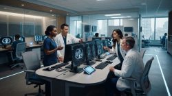
Diagnostic workstation software serves as the primary interface for radiologists to interpret, manipulate, and report on medical imaging. It’s the central hub where clinical data transforms into actionable insights. In the past, this meant sitting at a dedicated terminal in a dark room. Today, the landscape has shifted toward software-defined, device-agnostic platforms that allow for high-stakes interpretation from almost anywhere. This evolution represents a significant relief for departments previously burdened by rigid, location-dependent workflows.
At its core, modern radiology viewing software acts as the sophisticated front-end for your Picture Archiving and Communication System (PACS). While the PACS handles the heavy lifting of storage and retrieval, the workstation software manages the user experience. Choosing the right interface directly impacts diagnostic speed and clinical accuracy. By streamlining how images are rendered and reports are generated, facilities can significantly reduce operational overhead and improve the throughput of their entire imaging department.
The Shift from Proprietary Hardware to Software-Defined Workstations
The era of the $50,000 hardware-locked workstation is ending. Historically, vendors forced hospitals into expensive, proprietary hardware bundles to ensure performance. Those days are gone. Modern clinics now prioritize scalability and fiscal responsibility by choosing software that runs on standard, high-performance medical displays. This transition relies heavily on the DICOM standard and HL7 protocols to ensure seamless communication between disparate systems. Standardizing on these protocols allows for a more flexible infrastructure, letting you add new seats or upgrade your processing power without being held hostage by a single hardware manufacturer’s lifecycle.
Regulatory Standards: FDA Cleared vs. General Use Viewers
It’s vital to distinguish between clinical review and diagnostic interpretation. General use viewers are excellent for referring physicians who need a quick look at a patient’s study. However, primary diagnosis requires a higher level of scrutiny. Diagnostic-grade radiology viewing software must carry FDA Class II clearance and CE labeling to guarantee pixel integrity and tool precision. These certifications ensure that the measurements you take and the densities you observe are clinically reliable. For 2026, diagnostic-grade software must conform to the NEMA PS 3.1 – 3.20 2025d version of the DICOM standard to meet the latest FDA declarations of conformity for primary interpretation.
Essential Clinical Features for Diagnostic-Grade Viewing
High-performance 2D rendering is the bedrock of any diagnostic environment. When a radiologist opens a study, the software must respond with instantaneous fluid manipulation. Tools like zoom, pan, and windowing shouldn’t just work; they must feel natural and lag-free. This responsiveness directly impacts diagnostic speed, especially when navigating large datasets from modern CT or MR modalities. A high-quality radiology viewing software also excels at study comparison, allowing clinicians to load current and prior examinations side-by-side. This capability is vital for tracking disease progression or assessing treatment efficacy in oncology and chronic care.
Efficiency doesn’t stop at 2D images. Multimodal support is a non-negotiable standard for 2026. A single interface should handle everything from CT and MR to PET, Mammography, and Ultrasound. This unified approach removes the friction of switching between different applications for different study types. Effective clinical outcomes rely on Integrating Workstations with RIS and EMR to provide a complete patient history alongside high-resolution images. Many clinics find that switching to a modern software-defined PACS eliminates the lag and high costs of legacy systems.
Advanced Manipulation and Reconstruction Tools
Modern workflows require more than just looking at slices. Multiplanar Reconstruction (MPR) is essential for visualizing anatomy across axial, sagittal, and coronal planes simultaneously. This allows for a 3D understanding of complex structures that a single plane cannot provide. For surgical planning, 3D volume rendering provides a clear map of pathology, helping surgeons visualize the approach before entering the operating room. In oncology, PET-CT fusion tools are critical. By aligning functional metabolic data with anatomical structures, clinicians can pinpoint the exact location of high-activity lesions with surgical precision.
Reporting and Measurement Accuracy
Diagnostic confidence rests on the precision of the tools provided. Radiologists need reliable measurement capabilities for lengths, angles, and Hounsfield units (HU) to quantify findings accurately. These tools must be easy to access and intuitive to use. Beyond the pixels, the software should integrate seamlessly with speech recognition and structured reporting modules to close the loop between interpretation and documentation. Display consistency is equally important. The software must ensure that image quality remains uniform across different monitor configurations, maintaining the integrity of the diagnostic interpretation regardless of the hardware being used.
The Rise of Zero-Footprint and Web-Based PACS Systems
A zero-footprint viewer represents a fundamental shift in how facilities deploy radiology viewing software. Unlike traditional “thick client” applications that require local installation on every workstation, these systems run entirely within a standard web browser. This means your IT department no longer needs to visit individual desks for updates or troubleshooting. Everything is managed centrally on the server. This centralized model provides immediate relief to overburdened IT teams, ensuring that every user always accesses the most current, secure version of the software without downtime.
Security remains the top priority in this transition. Managing HIPAA compliance in a cloud-based environment requires robust encryption and strict access controls. Modern web-based PACS systems leverage secure streaming protocols that never store sensitive patient data on the local device. This architecture significantly reduces the risk of data breaches associated with lost or stolen hardware. By moving to a software-defined ecosystem, facilities can maintain a high security posture while enjoying the flexibility of a modern, integrated platform.
Teleradiology and Remote Reading Flexibility
Radiologists now demand the ability to work securely from any location. Web-based systems empower this flexibility by removing the need for specialized, proprietary hardware at home offices. The challenge has always been latency; however, modern streaming protocols have solved this. These systems use “just-in-time” delivery to ensure lightning-fast image loading, even over standard internet connections. The software intelligently prioritizes the images you need most, maintaining diagnostic quality even when bandwidth conditions fluctuate. It’s a pragmatic solution that allows for seamless teleradiology without compromising clinical standards.
SMAART-SHARE: Cloud-Based Collaboration
The days of burning physical CDs are numbered. SMAART-SHARE provides a secure, cloud-based alternative that allows for instant image sharing between patients and providers. This shift has a massive fiscal impact, eliminating the costs of CD burners, physical media, and the administrative time spent managing them. Beyond simple sharing, SMAART-SHARE enhances collaborative viewing during multidisciplinary team meetings. Multiple clinicians can view and discuss the same diagnostic-grade images in real time, regardless of their physical location. This level of seamless integration turns the radiology viewing software into a truly collaborative imaging ecosystem, reducing operational friction and improving the speed of care.

Interoperability: Integrating Workstations with RIS and EMR
Viewing images in a vacuum is a clinical risk. A siloed viewer creates a massive bottleneck for the entire department, forcing radiologists to interpret studies without the full patient context. Radiologists need more than just high-resolution pixels; they need the patient’s clinical history, lab results, and previous reports. Integrating your radiology viewing software with your Electronic Medical Record (EMR) and Radiology Information System (RIS) ensures that every study is accompanied by the data needed for an accurate diagnosis. This connectivity reduces errors and provides the “why” behind the findings in every image. Understanding the benefits of integrated RIS and EMR is essential for any facility looking to eliminate the login fatigue and manual data entry that plague disconnected infrastructures.
EMR and HIS connectivity means embedding imaging links directly into the patient’s electronic record. Clinicians shouldn’t have to hunt for studies across different platforms. When the system is integrated, a single click from the EMR patient profile launches the diagnostic viewer. This synergy syncs imaging with scheduling, billing, and the broader clinical history. It creates a unified patient record that serves the entire facility, offering a level of modern efficiency that legacy systems simply can’t match.
The RIS-PACS Synergy: SMAART-RIS and SMAART-PACS
Integrated systems eliminate the headache of duplicate data entry and manual errors. When you use SMAART-RIS alongside SMAART-PACS, data flows naturally from the initial order to the final report distribution. We design our tools to streamline the integration of SMAART-RIS with existing HIS platforms, ensuring your team spends less time on administrative tasks and more on patient care. This unified approach offers immediate relief from the operational friction of disconnected tools. You can experience the benefits of a truly integrated ecosystem by upgrading to SMAART’s RIS and PACS solutions.
HL7 and DICOM: The Languages of Interoperability
Interoperability depends on two primary languages: HL7 and DICOM. HL7 handles the transmission of patient demographics and order information, while DICOM manages the images themselves. It’s critical that your workstation software can “talk” to third-party EMRs without friction. Using open standards is the best way to future-proof your facility against vendor lock-in. It gives you the freedom to choose the best tools for your clinic without worrying about whether they’ll communicate with your existing infrastructure. Modern radiology viewing software must be an enterprise integration point, not just a standalone viewer. Facilities that have struggled with interfacing PACS with EMR across disparate systems know firsthand how critical open-standard compliance is to achieving a truly unified clinical workflow.
Future-Proofing Your Facility with SMAART Medical Systems
Modernizing your imaging department shouldn’t feel like a financial gamble. Many facilities remain stuck with legacy systems because the cost of a total overhaul seems insurmountable. SMAART Medical Systems, Inc. offers a pragmatic path forward. We provide affordable, full-feature solutions designed specifically for the unique pressures of hospitals and independent clinics. Our suite of products, including SMAART-PACS and SMAART-RIS, acts as a modernizer for your facility. We focus on seamless integration with your existing infrastructure to minimize operational friction from day one.
Growth requires flexibility. That’s why our licensing models are built to be scalable, adapting as your patient volume increases or your facility expands. We don’t believe in one-time transactions. Instead, we offer comprehensive support and a long-term partnership. Our team is deeply invested in your ongoing success, providing the technical authority and reassuring warmth you need to thrive in a competitive healthcare landscape. Choosing the right radiology viewing software is a strategic decision that impacts your clinical outcomes and your bottom line for years to come.
Reducing Radiology Overhead with SMAART-PACS
High-maintenance hardware is a significant drain on facility resources. SMAART-PACS shifts the focus from proprietary terminals to high-performance radiology viewing software that runs on standard medical-grade displays. This software-defined approach reduces your dependency on expensive, hardware-locked systems that require frequent, costly repairs. By integrating with the technology you already have, we help you achieve modern efficiency without the “rip and replace” price tag. Before committing to any upgrade, healthcare leaders should consider calculating ROI for a new PACS system to build a clear financial case that accounts for total cost of ownership and long-term operational savings. SMAART Medical Systems, Inc. provides the professional confidence needed to navigate complex radiology management with ease.
Next Steps: Evaluating Your Current Workflow
The first step toward relief is identifying where your current system is failing you. Conduct a digital audit of your imaging department to pinpoint specific bottlenecks. Are you losing time to slow image loading? Is your team still struggling with physical CDs? If so, it’s time to explore the advantages of cloud-based sharing with SMAART-SHARE. This tool replaces outdated physical media with a secure, collaborative ecosystem that connects providers and patients instantly. Moving your workflow to the cloud reduces IT overhead and ensures your data is accessible whenever and wherever it’s needed.
Transitioning to modern imaging tools is about more than just better pixels. It’s about reclaiming your time and refocusing your budget on patient care. Experience a more streamlined workflow – request a demo of SMAART-PACS today.
Embrace the Future of Integrated Imaging
The transition to software-defined, device-agnostic platforms marks the end of the proprietary hardware era. We’ve explored how modern radiology viewing software now serves as an enterprise integration point, bridging the gap between high-end clinical tools and administrative efficiency. By prioritizing interoperability and adopting zero-footprint architectures, your facility can finally eliminate the bottlenecks of siloed data. This shift provides immediate relief from operational strain, allowing your team to focus on diagnostic precision rather than technical workarounds.
SMAART Medical Systems delivers the pragmatic tools needed to achieve this modernization with fiscal responsibility. Our integrated RIS-PACS solutions and affordable cloud-based sharing through SMAART-SHARE ensure your department remains agile and connected. With seamless EMR integration, you can build a truly unified patient record that supports better clinical outcomes across your entire organization. It’s time to move past the limitations of legacy systems and embrace a partner dedicated to your ongoing success.
Upgrade your imaging workflow with SMAART-PACS today to experience a more efficient, integrated future. Your department deserves a system that works as hard as you do.
Frequently Asked Questions
What is the difference between a DICOM viewer and a diagnostic workstation?
A DICOM viewer is a general purpose tool used for clinical review by referring physicians. In contrast, a diagnostic workstation is an FDA cleared environment designed for primary interpretation. These workstations include advanced toolsets like MPR and 3D reconstruction. They must meet strict regulatory standards to ensure that the measurements and densities observed by the radiologist are clinically reliable for making a final diagnosis.
Is zero-footprint PACS software safe for diagnostic use?
Yes, modern zero-footprint radiology viewing software is safe for diagnostic use if it carries FDA Class II clearance. These systems use secure streaming protocols to deliver pixel-perfect images directly to a web browser without local installation. This architecture maintains high clinical standards while offering the flexibility of remote access. It allows radiologists to interpret studies from almost any location without compromising image integrity or patient data security.
Can diagnostic workstation software integrate with my existing EMR?
Modern workstations use HL7 and DICOM protocols to integrate seamlessly with existing EMR and HIS platforms. This connectivity allows clinicians to launch studies directly from the patient record with a single click. It ensures the radiologist has full clinical context, such as lab results and previous reports, alongside the images. This synergy reduces diagnostic errors and removes the operational friction of switching between disconnected software applications.
Does medical imaging software require specialized hardware in 2026?
No, the industry has shifted toward software-defined solutions that run on standard, medical-grade displays. While you still need high-resolution monitors for diagnostic accuracy, you’re no longer locked into proprietary, vendor-specific hardware terminals. This change provides significant relief from the high costs of legacy systems. It allows facilities to upgrade their processing power or add new seats using standard professional hardware, greatly simplifying long-term IT maintenance.
How does cloud-based image sharing improve patient care?
Cloud-based sharing through tools like SMAART-SHARE improves care by providing instant, secure access to images for both patients and providers. It eliminates the delays and administrative headaches associated with burning physical CDs. Collaborative viewing features allow multidisciplinary teams to discuss cases in real time from different locations. This speed leads to faster treatment decisions and a more integrated, responsive experience for the patient throughout their care journey.
What are the minimum hardware requirements for diagnostic-grade viewing?
Minimum requirements typically include a high-resolution medical monitor, such as a 3MP or 5MP display, and a workstation with a dedicated GPU. While the radiology viewing software itself is device-agnostic, the display must meet specific brightness and contrast standards for primary diagnosis. A stable, high-speed internet connection is also essential for fluid image manipulation in web-based environments, ensuring that tools like zoom and pan remain lag-free during interpretation.
Is SMAART-PACS HIPAA compliant?
Yes, SMAART-PACS is fully HIPAA compliant and includes robust encryption and strict user access controls to protect sensitive data. The system is designed with security as a core priority, ensuring that protected health information is encrypted both at rest and during transmission. This provides administrative relief by meeting all regulatory requirements for data privacy. It allows your facility to focus on clinical excellence while maintaining a high security posture.
How much can a facility save by switching to a software-defined workstation?
Facilities see substantial savings by eliminating proprietary hardware maintenance and reducing local IT overhead. By moving to a system like SMAART-PACS, you avoid the recurring costs of hardware-locked terminals that require specialized service. Additional savings come from centralized software updates and the removal of physical media costs through cloud-based sharing. This pragmatic approach to imaging allows you to reallocate your budget toward clinical staff and improved patient services.